Retrognathia
Now we have inferior growth of the lower jaw. Because the lower jaw is receding, the upper jaw appears to be protruding. Patients often express that they are concerned about the protruding part, but the problem is the lower jaw receding. Although it is before the accelerated growth period, growth is deeply involved, so it is necessary to examine and diagnose including the TMJ. Especially for the TMJ, an MRI test is performed to understand the state of disc displacement.
In this case, early stage of disc displacement was found. We want to induce maximum growth of the lower jaw during the accelerated growth period. To prepare for this, we first applied a splint to the lower jaw to stabilize the position of the jaw, since we found instability in the position of the upper and lower jaw and CBCT images of the TMJ showed that the mandibular head had shifted significantly from the glenoid fossa. Then, to correct the vertical relationship between the molars and premolars, minimal orthodontic appliances and headgear were placed on the molars.
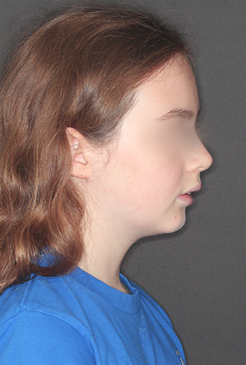
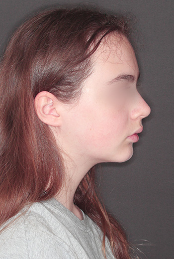
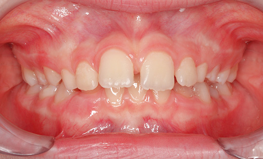
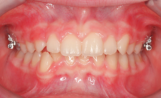
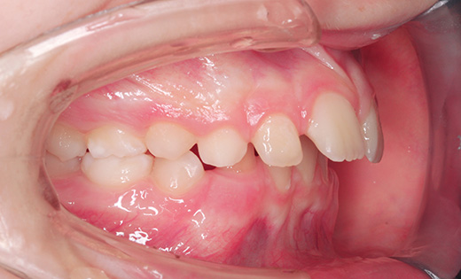
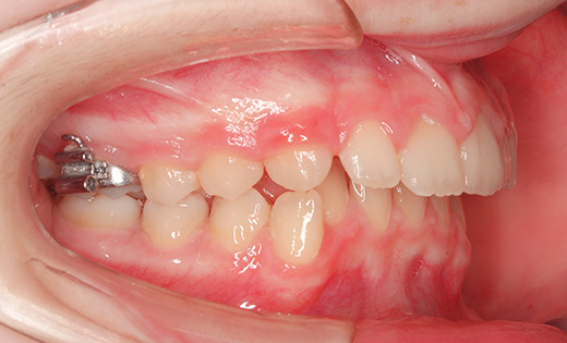
The left is a photo of the profile and the inside of the mouth before treatment, and the right is a photo of the post-treatment. At the initial visit, the patient's lower jaw was significantly misaligned at the temporomandibular joint. The original bite at the initial examination would have resulted in the lower jaw being further receded, and the chin would have been further receded in the profile.
We placed a splint on the lower jaw, minimal orthodontic appliances on the upper jaw, and headgear on the molars. Stabilizing the lower jaw is essential to maximize the growth of the lower jaw in preparation for the upcoming accelerated growth period. The line from the nose down to the upper lip, lower lip, and chin has been neatly aligned.
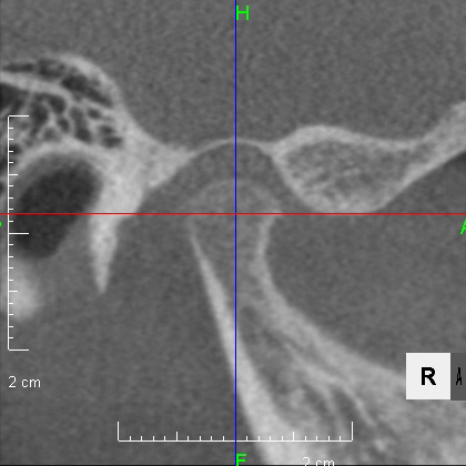
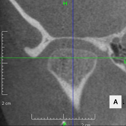
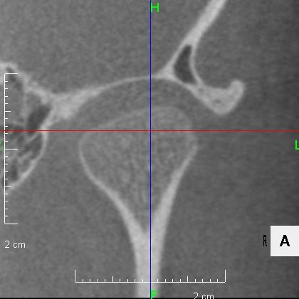


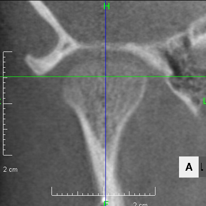
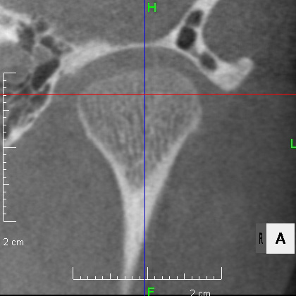
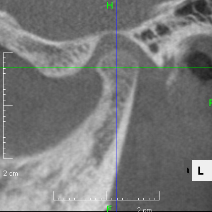
CBCT images of the TMJ before orthodontic treatment at the top and after orthodontic treatment at the bottom.
Patients bite in a place that is easy for them to bite, so if we do not observe carefully, we may miss the correct relationship bite. As you can see from the photo above, before treatment the bite was shifted, which caused the left and right mandibular condyles to shift forward and downward away from the glenoid fossa, putting strain on the TMJ. By wearing a splint, the mandibular condyles were placed in their natural position and stabilized. This is the position that puts the least strain on them.
The appliances included a splint on the lower jaw to stabilize its position, orthodontic appliances on the four upper front teeth and headgear on the molars to improve the position of the front teeth and to control the vertical height of the molars.
The treatment period was one year and six months, and the treatment cost was 350,000 yen (excluding tax, at that time).
- The commonly cited treatment risks at this time are the development of caries, gingivitis, and temporomandibular joint disorder, but none of these occurred in this case.
To summarize this case, the main focus of treatment was to prepare the TMJ for the upcoming period of accelerated growth and to bring out the growth of the lower jaw as much as possible. The key is the diagnosis and treatment procedure.