We have discussed the close relationship between the alignment of the teeth, the temporomandibular joint(TMJ), and the position of the lower jaw, as well as its effect on the face. In addition, we have explained that during the growth period, the TMJ is greatly involved in the growth of the lower jaw, so we do not move the teeth unnecessarily, but use a splint to stabilize the jaw as needed to promote growth. However, if there is an abnormality in the positional relationship between the upper and lower jaw, there is a need for intervention during the early growth period (around 6-9 years of age). When is intervention necessary? I will discuss next.
Underbite
Normal front teeth are placed with the upper front teeth in front of the lower front teeth, but when this is reversed with the lower front teeth placed in front of the upper front teeth, we call this “Underbite”.
There are two types of problems: one is simply a problem with the position of the teeth, and the other is a problem with the skeletal position of the upper and lower jaws. The latter skeletal pattern problem should be treated as early as possible because then, there is higher chance of positive changes in the growth pattern that occurs later in life.
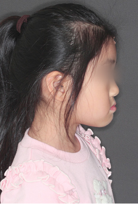
Look at the before and after pictures of the treatment below. Before treatment the lower front teeth are forward covering the upper front teeth. At first glance, it appears that the lower jaw is forward, but a closer look at the face reveals that the upper jaw appears to be receding and underdeveloped rather than the lower jaw being significantly forward. The lower jaw is protruding because of the retraction of the nasal wings. The upper and lower jaws grow at different times; the upper jaw growth ends around age 12, while the lower jaw growth continues thereafter. The time when the upper jaw growth can be induced will end early, so we need to hurry in this case.
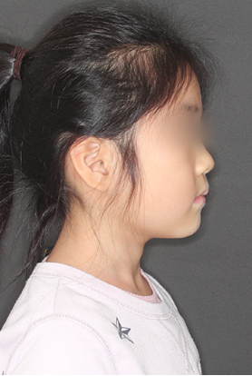
Left is a profile view before treatment and right is a profile view after treatment. The upper jaw was induced to grow forward. As for the relationship between the front teeth, the upper jaw itself was brought forward, so the teeth was not moved, but the upper front teeth was brought forward in relation to the lower front teeth.
The sides of the nose were pulled forward and the cheeks became puffy. The tip of the nose has also been brought forward, giving the face a more childlike appearance.
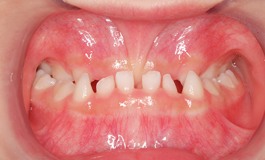
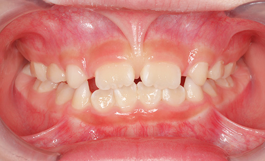
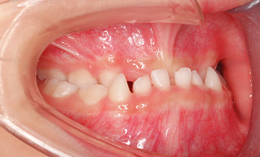
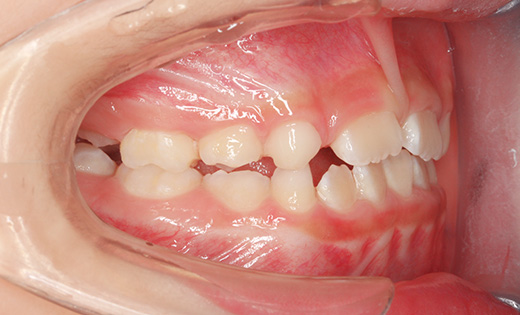
Intraoral photographs before treatment on the left and after treatment on the right. Before treatment, the lower front teeth are forward covering the upper front teeth. When the upper jaw is induced to grow forward, the upper front teeth are brought forward, although no tooth movement is performed in relation to the front teeth.
Before treatment, the lower front teeth are forward covering the upper front teeth. This relationship continues to the back teeth, and the molars also have a crossbite with the lower molars on the outside of the upper molars. Even during the primary dentition period, there can be problems with the TMJ. An examination including the TMJ is necessary even during this period. Since the upper jaw is narrow, the upper jaw was expanded laterally, the bony junction of the upper jaw was loosened and the upper jaw was pulled forward. The front teeth are not moved, but the upper front teeth are brought forward. The sides of the nasal wings have moved forward and the cheeks are puffier. The tip of the nose has also been brought forward, giving the face a childlike appearance.
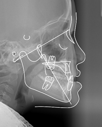
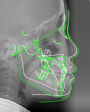
Comparison of standard head X-ray photographs before treatment on the left and after treatment on the right. The upper jaw was induced to grow forward. Before treatment, the toothless elderly-looking profile was greatly changed to a plump, child-like face.
At the time of initial examination, moderate deformity was already observed in the left temporomandibular joint and the patient had a bite that placed a burden on the temporomandibular joint, so we first performed splint therapy and confirmed improvement of the mandibular head deformity with CBCT before proceeding to correct the skeletal structure.
The upper jaw was placed with a rapid expansion device (requires rapid expansion) and a face mask to pull the jaw forward. The lower jaw was fitted with a splint to protect the temporomandibular joint from rapid changes in occlusion. The treatment period was one year and nine months, and the treatment cost was 350,000 yen (excluding tax, at that time).
- The occurrence of caries, gingivitis, and TMJ disorder are often cited as common treatment risks during this period, but in this case, the bone deformity of the TMJ was corrected.
①
②
③
④
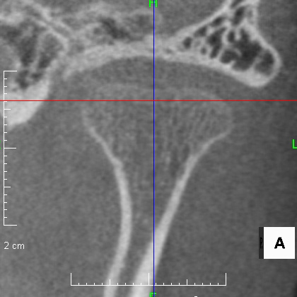
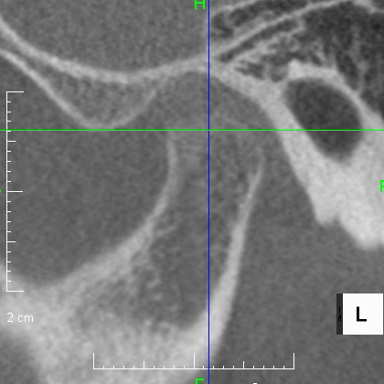
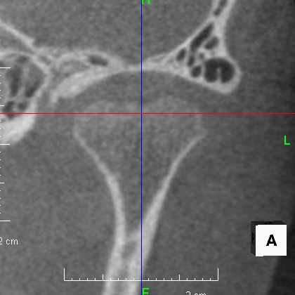
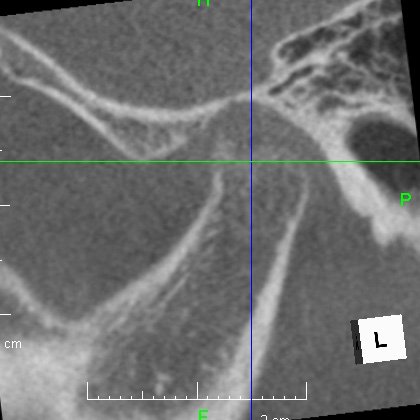
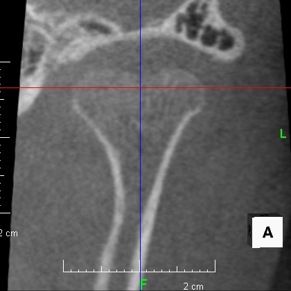
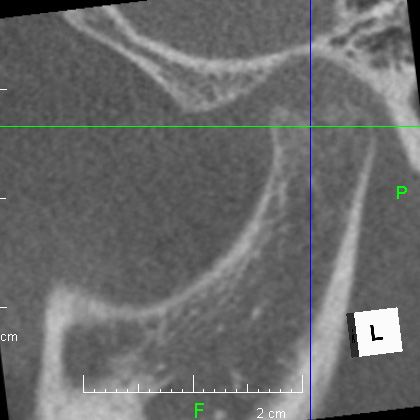
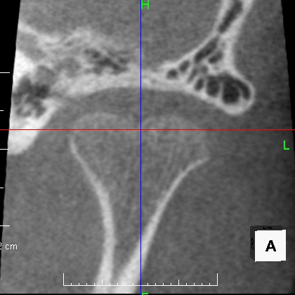
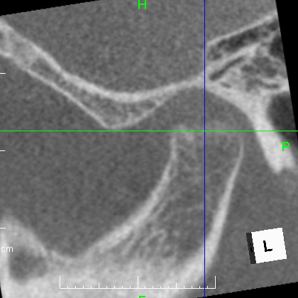
Comparison of CBCT images of the left temporomandibular joint. (1) At the time of initial examination (age 6 years and 2 months), a resorption image was seen in the upper part of the mandibular head. The surface is rough and concave. (2) About 5 months after the patient started to wear the splint on the lower jaw, the roughness of the resorption image became a little smoother. (3) At the end of the initial treatment (8 years and 3 months old), the surface became smoother. (4) Two years after the treatment (age 10 years and 3 months), she continues to wear the splint. The problem has almost recovered as if there was no problem at all. It is important to emphasize that in this case, the MRI showed that the disc displacement had not progressed that far, so we were able to miraculously obtain this kind of restoration. All thanks to the early treatment.
To summarize this case, the timing of orthodontic treatment varies from case to case, and early action is essential if the skeletal pattern is problematic. If the patient can be treated at this time, growth can be induced and treatment possibilities can be expanded. Although not visible on the surface, there was already moderate deformity of the left temporomandibular joint. Although most of the attention is focused on the upper jaw, we are also watching for the normal growth of the lower jaw. Regardless of the timing of orthodontic treatment, we will examine and diagnose the patient's situation including the temporomandibular joint, fully understand the patient's situation, and provide appropriate treatment that can only be done at the appropriate time, and although orthodontic treatment to align the teeth is often necessary in the future, until then we focus on watching the growth of the jaw.